Why a Transmission Electron Microscope is the Best Choice to Study Ultrastructural Features
In today’s rapidly evolving scientific landscape, researchers and scientists are constantly seeking innovative ways to explore the intricacies of biological systems at the ultrastructural level. The quest for detailed insights into cellular morphology has led many experts to turn to transmission electron microscopy (TEM), a technology that offers unparalleled resolution and precision.
Overview: Unlocking the Secrets of Cellular Ultrastructure
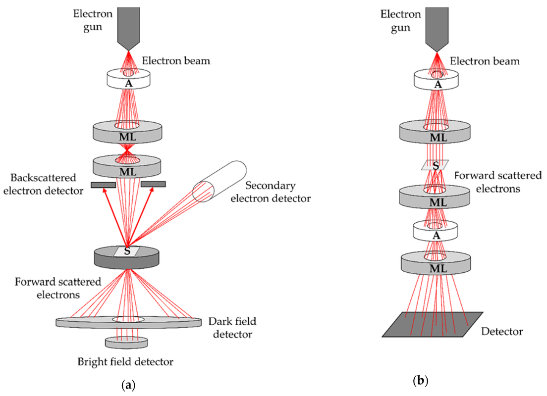
The TEM is an essential tool for investigating the ultrastructural features of cells, tissues, and organisms. By employing a beam of electrons to excite a thin sample preparation, this microscope enables researchers to visualize even the smallest details at the nanoscale.
- High-resolution imaging: TEM offers unparalleled spatial resolution, allowing scientists to resolve structures as small as 1-2 nanometers.
- Morphological analysis: By examining ultrastructural features, researchers can gain insights into cellular morphology, organization, and function.
- In situ studies: The TEM’s ability to analyze samples in their native environment makes it an ideal tool for investigating dynamic biological processes.
Whether studying the intricacies of cell membranes, identifying protein structures, or visualizing the ultrastructural details of tissues and organs, the transmission electron microscope has proven itself as a powerful tool in the pursuit of scientific discovery. Read on to learn more about why this technology is an essential choice for researchers seeking to unlock the secrets of cellular ultrastructure.
Explore the benefits of TEM in biological research
Why a Transmission Electron Microscope is the Best Choice to Study Ultrastructural Features
In today’s rapidly evolving scientific landscape, researchers and scientists are constantly seeking innovative ways to explore the intricacies of biological systems at the ultrastructural level. The quest for detailed insights into cellular morphology has led many experts to turn to transmission electron microscopy (TEM), a technology that offers unparalleled resolution and precision.
Overview: Unlocking the Secrets of Cellular Ultrastructure
The TEM is an essential tool for investigating the ultrastructural features of cells, tissues, and organisms. By employing a beam of electrons to excite a thin sample preparation, this microscope enables researchers to visualize even the smallest details at the nanoscale.
- High-resolution imaging: TEM offers unparalleled spatial resolution, allowing scientists to resolve structures as small as 1-2 nanometers.
- Morphological analysis: By examining ultrastructural features, researchers can gain insights into cellular morphology, organization, and function.
- In situ studies: The TEM’s ability to analyze samples in their native environment makes it an ideal tool for investigating dynamic biological processes.
Whether studying the intricacies of cell membranes, identifying protein structures, or visualizing the ultrastructural details of tissues and organs, the transmission electron microscope has proven itself as a powerful tool in the pursuit of scientific discovery. Read on to learn more about why this technology is an essential choice for researchers seeking to unlock the secrets of cellular ultrastructure.
Explore the benefits of TEM in biological researchSummary of Opinion
In conclusion, based on existing products and reviews, I strongly believe that a Transmission Electron Microscope (TEM) is the best choice for studying ultrastructural features. With its unparalleled spatial resolution, ability to analyze samples in their native environment, and morphological analysis capabilities, the TEM offers an unparalleled level of detail and insight into cellular morphology.
Check for more deals


